


Bone Anatomy and Function
Overview
- Bone is a living tissue with its own blood vessels, living cells proteins and minerals
- It is the hardest living tissue in the body
- Largest bone in human body is the femur (tibia is second)
- Smallest bone in human body is the stapes in the middle ear which is just 3mm long
- >99% of the body’s calcium is within the bones and teeth
- Is made up of two components:
- Organic component:
- 40% of dry weight
- Numerous proteins:
- Collagen:
- Makes up 90% of organic component
- Primarily type I collagen
- Provides tensile strength (resistance to tension)
- Proteoglycans:
- Provides strength to compression
- Inorganic component:
- 60% of dry weight
- Primary mineral is calcium hydroxyapatite:
- Provides main strength to compression
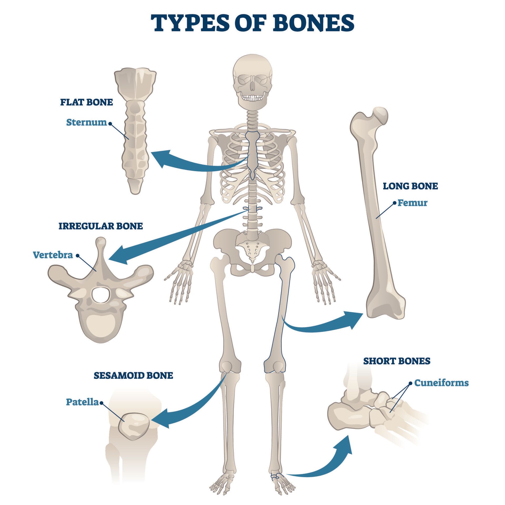
What are the different types of bones?
- Long bones:
- Located in the limbs and are longer than they are wide:
- Upper limb:
- Composed of shoulder, arm, elbow, forearm, wrist, hand
- Long bones are humerus of the arm, radius and ulna of the forearm, metacarpals and phalanges of the hand
- Lower limb:
- Composed of hip, thigh, knee, leg, ankle, foot
- Long bones are: femur of the thigh, tibia and fibula of the leg, metatarsals and phalanges of the foot
- Short bones:
- Length and width are similar
- Roughly cube shaped
- Provide stability and movement
- Located in wrist and ankle joints
- Flat bones:
- Flat and wide
- Skull
- Sternum
- Ribs
- Irregular bones
- Vertebrae:
- They are the bones of the spinal column
- Pelvis:
- Made up of the pubis, ilium and ischium
- Sesamoid bones:
- They are bones found within a tendon or muscle that glide over joints
- They are named so because they resemble a sesame seed
- Their function is to:
- Reduce friction on the tendon as it glides over bony prominence
- Provide mechanical advantage by increasing the lever arm of the pull of the tendon:
- That means that they amplify the force created by the muscle
- The patella for instance amplifies the pull of the quadriceps by 30-40%
- Examples of sesamoid bones:
- Patella:
- The largest sesamoid bone in the human body
- In the flexor tendons of the big toe and thumb
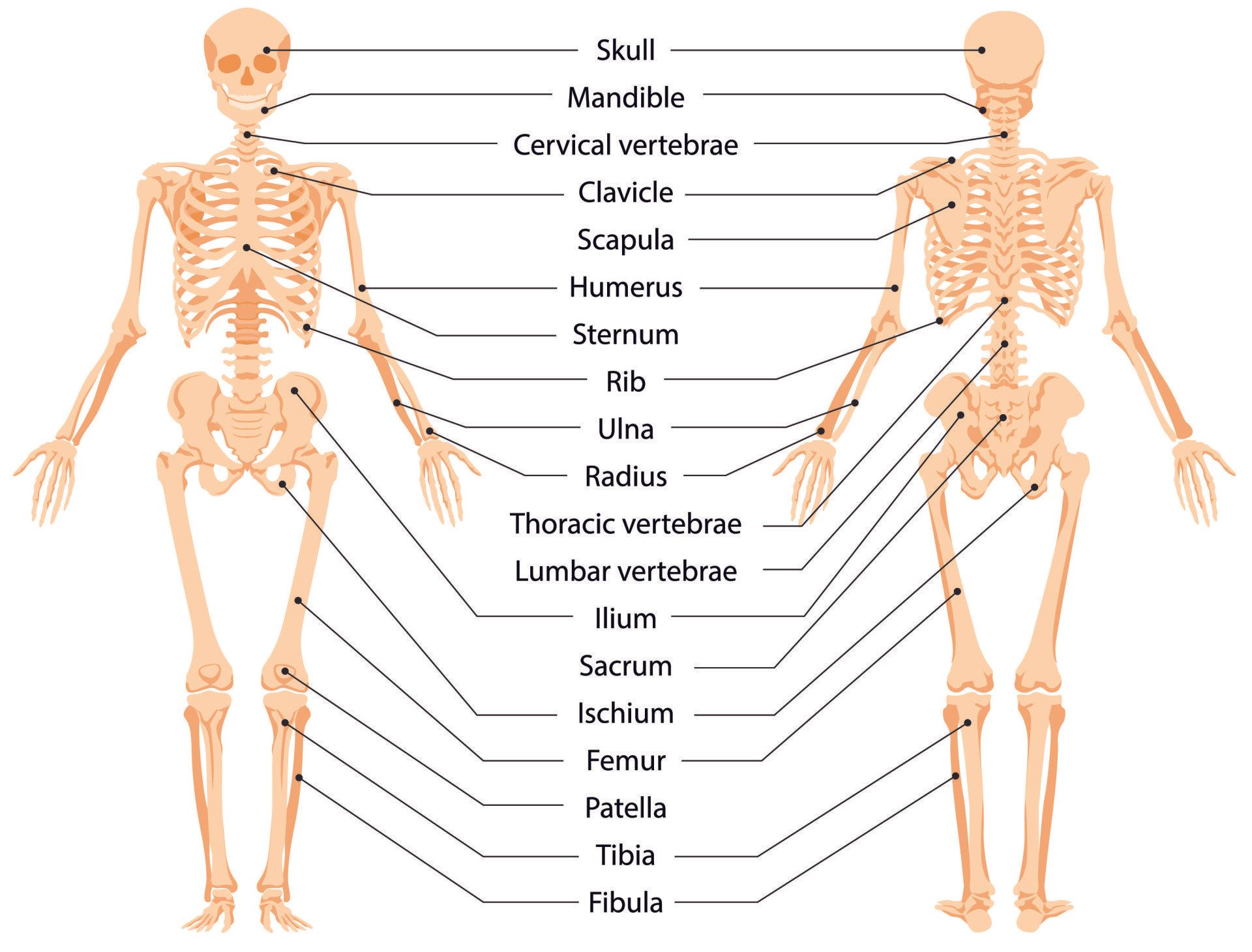
How many bones are there in the human skeleton?
- We are born with around 300 soft bones
- As we grow these soft bones are ossified with deposition of minerals (calcium and phosphorus) and many of them fuse together
- There are 206 bones in the adult human skeleton which make up 15% of the body weight
- This excludes teeth and sesamoid bones which are found in tendons going over bony prominences
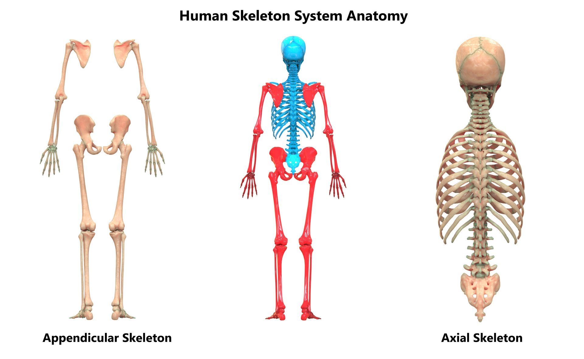
- The human skeleton can be subdivided into:
- Axial skeleton:
- 80 axial bones
- Located in the central axis of the body
- Skull, vertebrae, ribs, sternum
- Appendicular skeleton:
- 126 appendicular bones
- Composed of the bones that form the limbs with their attachment points to the axial skeleton called girdles:
- Limbs:
- Upper limbs
- Lower limbs
- Girdles – they are the attachment points for the limbs:
- Pelvic girdle:
- Attachment point for the femur (thigh bone) of the lower limb
- Consists of the ilium, ischium and pubis
- Pectoral girdle:
- Attachment point for the humerus (arm) of the upper limb
- Consists of clavicle and scapula
How do bones form?
- Bone formation (osteogenesis or ossification) begins in the 2nd month of foetal life and is completed in late adolescence
- The clavicle (collar bone) is the first bone to ossify in the developing embryo (~5th week of gestation)
- The clavicle is also the last to fully fuse – most happen by 20 years but can take as long as 25 years
- It occurs via two processes:
- Intramembranous ossification:
- Connective tissue membrane that is initially laid out is replaced by bone tissue
- Occurs in the formation of flat bones e.g. skull and clavicle
- Unlike endochondral ossification cartilage is not present during intramembranous ossification
- Endochondral ossification:
- Cartilage that is initially laid out is replaced by bone tissue
- Occurs in the formation of long bones (e.g. femur and tibia) and most other bones
- Ossification occurs at two centres:
- Primary ossification centre:
- Is the first area of bone that starts ossifying
- Usually appears before the baby is born
- Located in the central part of each developing bone
- In long bones this would be located in the centre of the diaphysis
- Secondary ossification centre:
- Appears after the primary ossification centre in the first years of life
- Located on either end of long bones in the epiphyses
- Longitudinal growth of a long bone occurs at the growth plate (physis):
- This is where new cartilage is formed continuously which is subsequently ossified
- The presence of cartilage means this is an area of weakness for the developing bone since cartilage is softer and weaker than bone
- They are seen as black lines across the width of long bones on X-rays and so can be confused as fractures
- When growth ceases at about 20 years the cartilage of growth plates is ossified and fuses with the rest of the bone
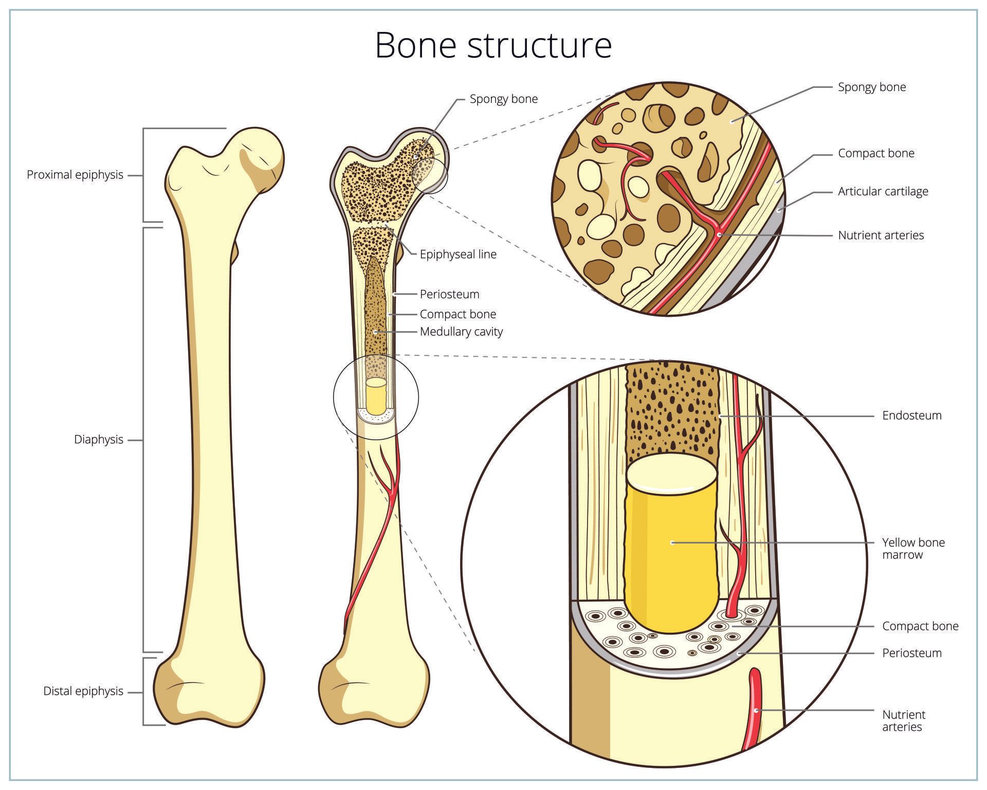
What are the different parts of a long bone?
- A long bone can be split into three sections:
- Diaphysis:
- Long slender middle portion of the bone also called the shaft
- The longest portion of the bone
- The cortex is thick and strong
- Metaphysis:
- Area between the diaphysis and epiphysis
- It contains the physis (growth plate):
- Location where the bone grows during childhood
- The physis is cartilaginous but is ossified once growth stops in early adulthood
- The bone becomes increasingly wider towards the epiphysis
- The cortex is thinner
- Has a rich bloody supply so prone to spread of infection from blood stream to the metaphysis leading to osteomyelitis
- Epiphysis:
- Large rounded end of the bone that forms the articular surface
- Section of bone from the joint surface to the physis
- Separated from the metaphysis by the physis


Structural organisation
- Cortical (compact) bone:
- Located on the periphery of the bone:
- If a bone is likened to a pipe, cortical bone is the wall of the pipe and cancellous bone is located inside the pipe
- Makes up 80% of skeleton by weight despite a much smaller proportion by volume than cancellous bone
- Strongest part of the bone but also more brittle
- Provides strength and protection to bones
- Slow turnover rate so remodels slowly
- Structure is highly organised into osteons:
- Cylindrical structures aligned along the lines of stress the bone is exposed to in order to maximise strength of the bone
- A fracture is a broken bone and is defined by discontinuity in the cortex of the bone and so state of the cancellous bone is irrelevant when considering whether a bone is fractured
- Cancellous (spongy) bone:
- Located inside the canal of the bone
- Less strong than cortical bone but more elastic and flexible
- High turnover rate so remodels quickly
- Less organised than cortical bone with a loose network of bone
- 30-90% of the cancellous bone is porous (has many holes) where bone marrow is located
- Despite making up the larger portion of bone by volume it only makes up 20% by weight
- During osteoporosis the porosity increases i.e. more and bigger holes so less bone per unit volume (density)


Types of bone cells
- Osteoprogenitor cells:
- Derived from mesenchymal stem cells
- Located in the bone marrow
- Produce osteoblasts
- Osteoblast:
- Forms new bone
- Stimulated by Vitamin D
- Regulates osteoclast activity
- Osteoclast:
- A large cell that reabsorbs existing bone and removes unwanted tissue
- Derived from haematopoietic stem cells
- Essential in bone remodelling such as due to stresses a bone is subjected to
- A target of many osteoporotic medications:
- By inhibiting their function less bone is eaten away thus increasing bone density
- Osteocyte:
- Mature bone cells
- Former osteoblasts trapped within the bone matrix they produce
- Commonest cell in bone ~90%
- Important in regulating the calcium and phosphorus concentrations
What are the functions of bones?
- Healthy bones are important throughout our lives
- Bones are living tissues and play a vital role in numerous functions:
- Support:
- They provide a framework for attachment of muscles via their tendons
- Ligaments connect bone to bone and so provide support across a joint
- Movement:
- As a result of this framework of muscles, tendons and ligaments, bones facilitate movement by acting as levers
- Joints act as the fulcrum (pivot point) across which the lever acts
- Protection:
- Bones protect vital organs:
- skull – brain
- rib cage – heart and lungs
- vertebrae – spinal cord
- pelvis – internal reproductive organs, bladder, lower digestive tract
- Mineral storage:
- Bones act as the storage bank for vital minerals such as calcium and phosphorus
- When they are in excess they can be stored in the bones
- When the body needs more of these minerals they can be released from the bones but beyond a point this will cause reduced bone mineral density resulting in increased fragility and increased risk of fracture
- Calcium is essential for muscle contraction and transmission of nerve impulses
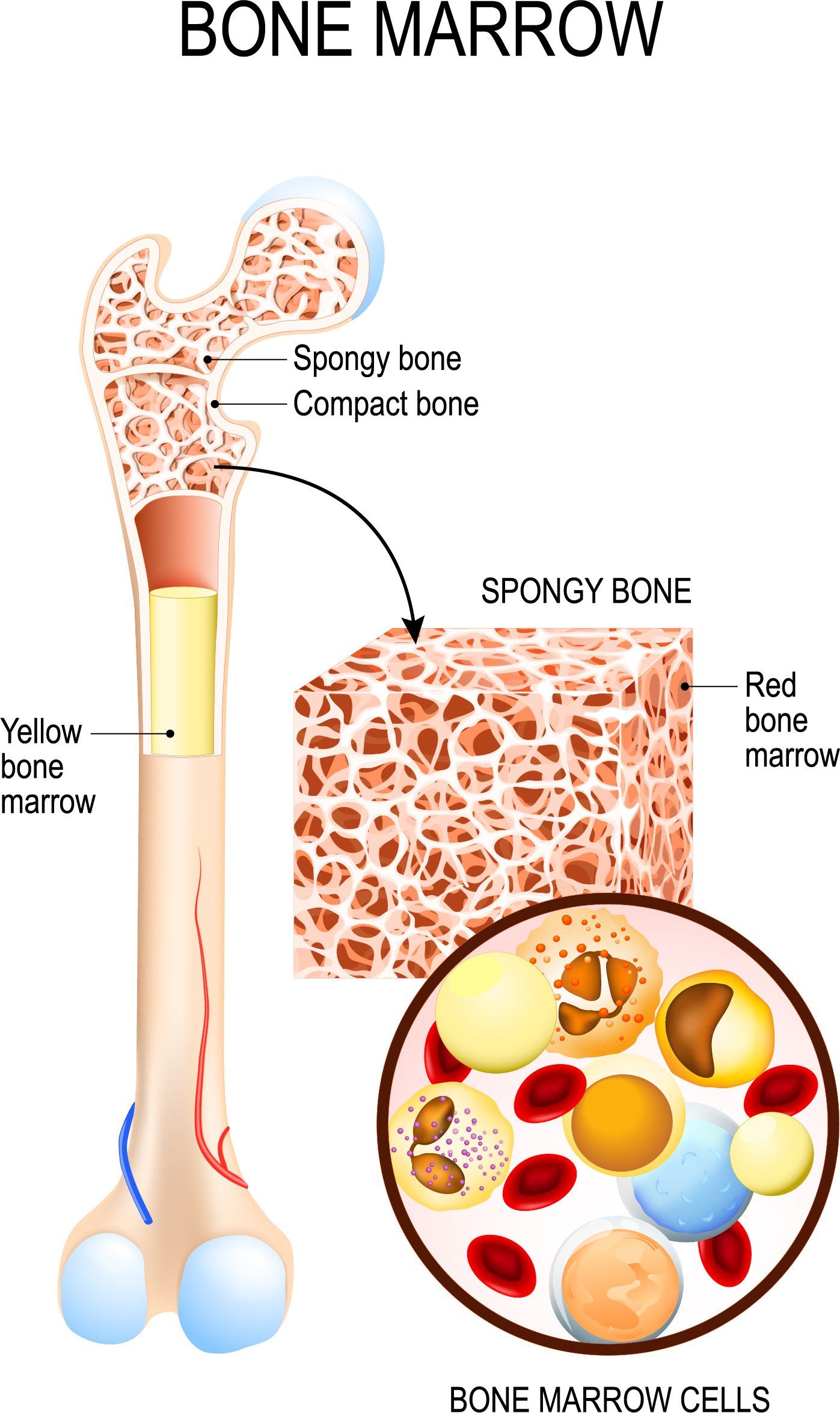
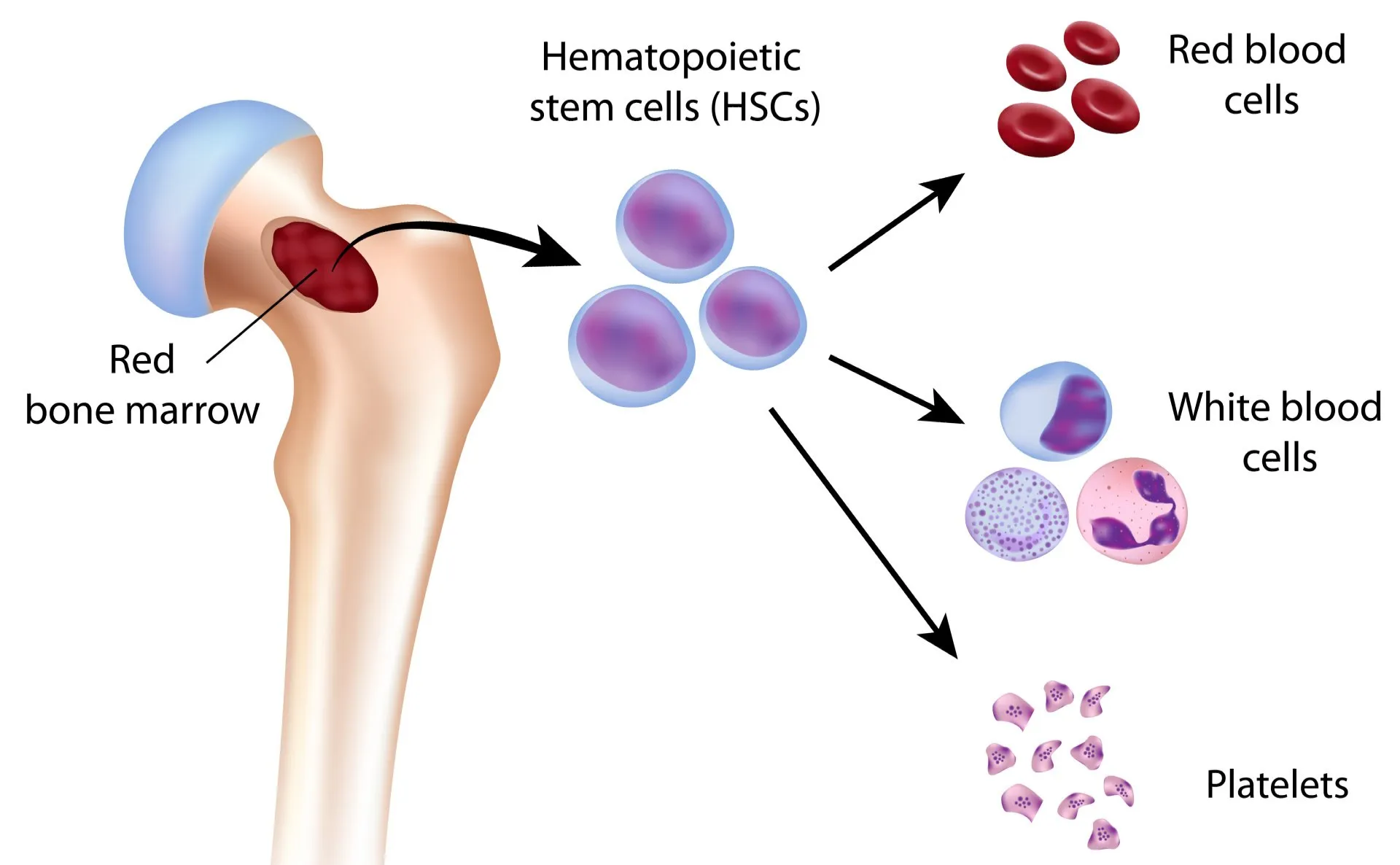
- Blood cell production:
- Red marrow is located between the cavities of a bone and is where the blood cells are formed by the multipotential haematopoietic stem cell:
- Red blood cells:
- They carry oxygen from the lungs to the rest of the body and return carbon dioxide from the body to the lungs
- White blood cells:
- They fight off infection
- Platelets:
- They help form blood clots in order to stop bleeding
- Energy storage:
- Lipids (fats) are stored within the yellow marrow of a bone
- Yellow marrow like red marrow, is also located within the cavities of a bone
- This fat can be broken down and used for energy when required


More relevant information can be found on:
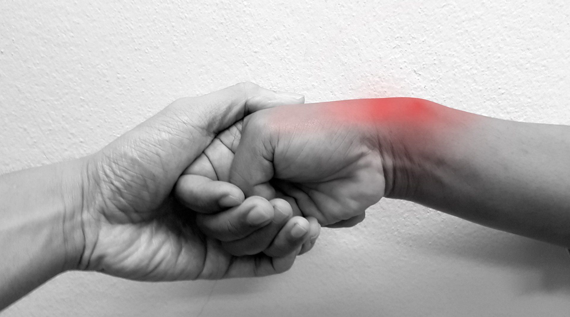
Introduction De Quervain’s tenosynovitis (also called De Quervain’s tendinosis) is a painful condition of two tendons at the level of the wrist on the side of the thumb It’s a repetitive strain injury due to overuse of two tendons used to move the thumb away from the other fingers
Introduction Cubital Tunnel Syndrome occurs when the ulnar nerve is compressed within a tunnel on the inner (medial) side of the elbow just behind the bony prominence of the inner aspect of the elbow called the medial epicondyle Cubital Tunnel Syndrome is the second most common cause of peripheral nerve compression: The most common one being carpal tunnel syndrome (compression of the median nerve at the wrist) The ulnar nerve is one of the three main nerves of the upper limb: The other two nerves of the upper limb are the median nerve and the radial nerve The ulnar nerve travels from the neck past the elbow and wrist and into the hand: Along the way it travels past some narrow areas where it can be constricted and cause symptoms for the patient The most common site of ulnar nerve compression is in the cubital tunnel at the elbow The second most common site is in Guyon’s canal in the hand When someone accidentally hits the inner side of the elbow (often termed hitting the funny bone) they get a sharp tingling sensation on the inner side of the elbow and forearm: This occurs because the ulnar nerve was hit at the site of the cubital tunnel where the nerve is close to the skin surface and therefore easily injured from outside forces
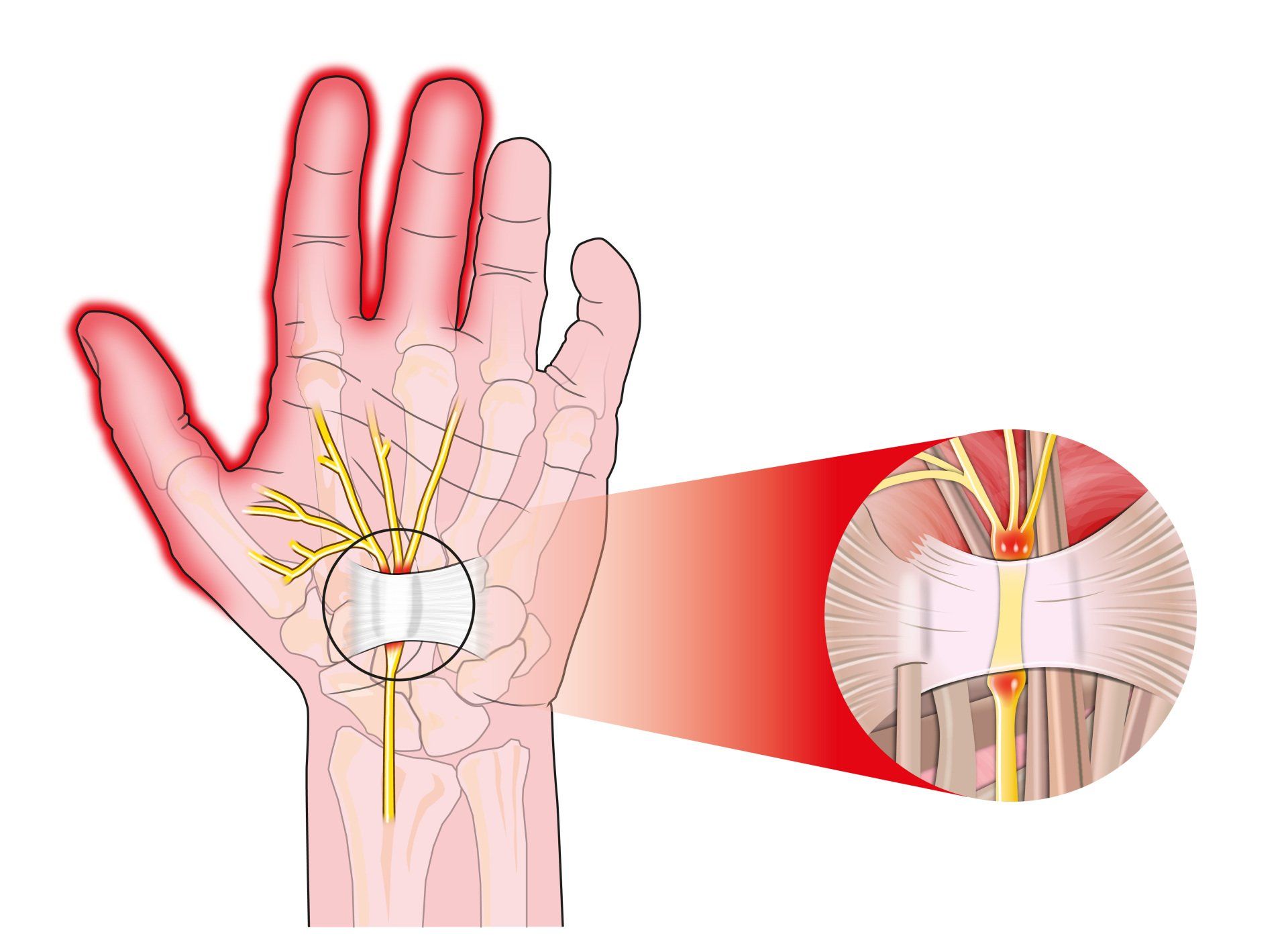
Introduction Carpal tunnel syndrome is a common condition that causes numbness, tingling and weakness in the hand specifically affecting the thumb, index and middle fingers: The little and ring fingers are not affected as they are supplied by another nerve called the ulnar nerve It is the commonest cause of peripheral nerve entrapment It is caused by compression of the median nerve as it passes from the forearm into the hand through a passage called carpal (i.e. wrist) tunnel The median nerve is one of three main nerves that supply the upper limb: The other two nerves are the ulnar nerve and the radial nerve
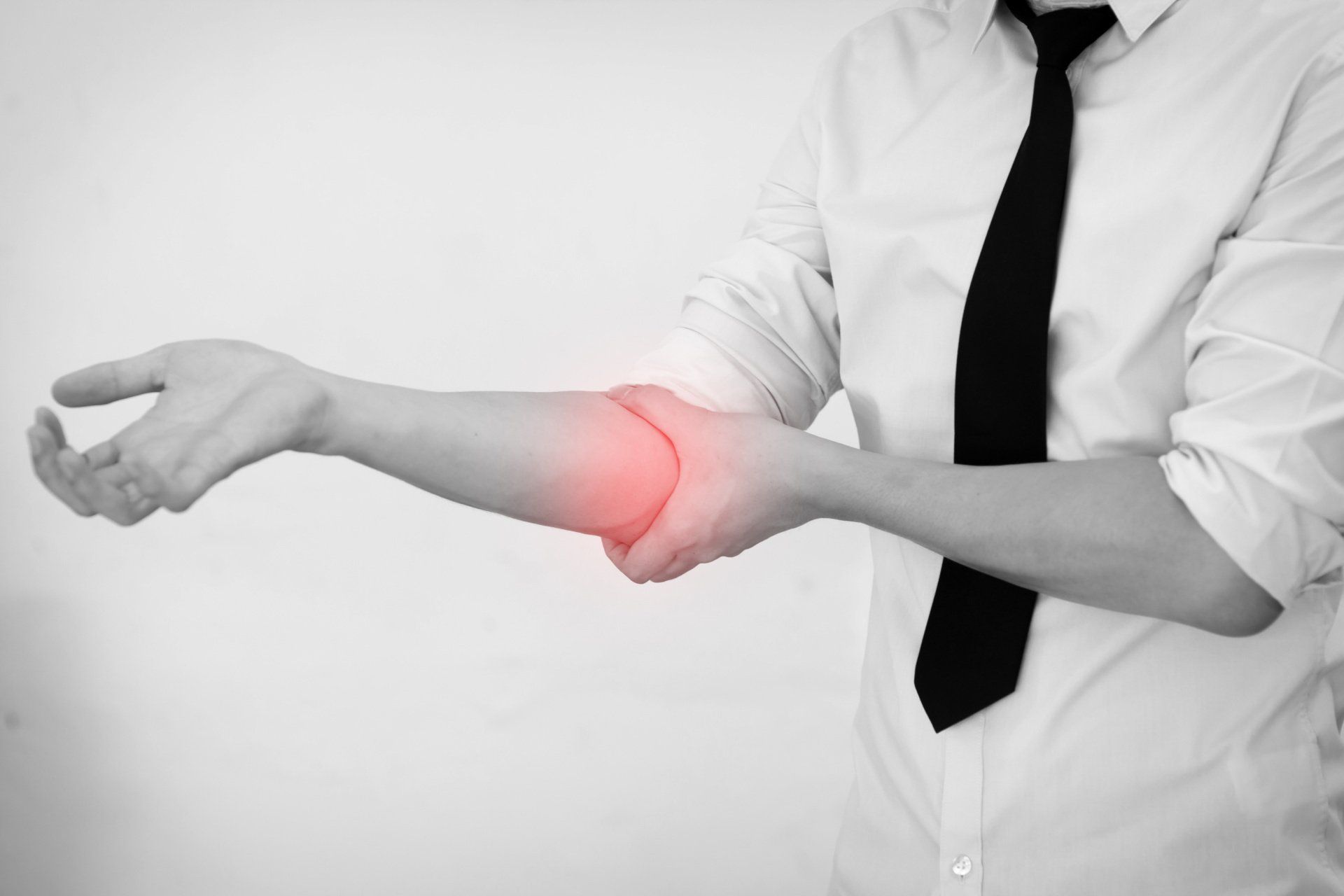
Introduction Tennis elbow (also known as lateral epicondylitis) is an overuse injury of the forearm tendons that originate over the lateral epicondyle of the humerus (bony prominence on the outside of the elbow) and act to bring the wrist backward away from the palm Whilst tennis players are particular prone to this condition it does not occur exclusively to them

Introduction This term is also known as repetitive motion or stress injury and occurs as a result of carrying out the same motion repeatedly over time causing injury to muscles and tendons It is associated with repetitive tasks, sustained or awkward position, forceful exertion, vibration or compressive forces It can affect almost any joint in the body Most commonly affected areas are hands, wrists, shoulders and neck It is thought to affect 5-10% of the general population but can be as high as 20-40% in specific working populations
The most common injuries in sailing, are in the waist (45%), knees (30%) and shoulders (20%). Causes of sailing injuries, windsurfing injuries, sprain or ruptured knee ligament. Injury prevention, protective clothing to avoid injuries in sailing.

Injuries such as concussion, anterior cruciate ligament injury, anterior gonalgia, fatigue fracture, tibial periostitis, ankle sprain, rotator cuff injury should be avoided for women to exercise.

Women have a 50% higher risk of developing pathologies when running and exercising. Prevention of Sport Injuries.

It is composed of three components: Low energy availability with or without eating disorder - Menstrual disorders - Decreased bone mineral density (BMD) - Treatment

What is the function of vitamin D - The benefits of vitamin D, what amount of vitamin D should I take, which foods are high in vitamin D, risks of vitamin D deficiency, the role of the sun, osteoporosis, causes, symptoms.